|
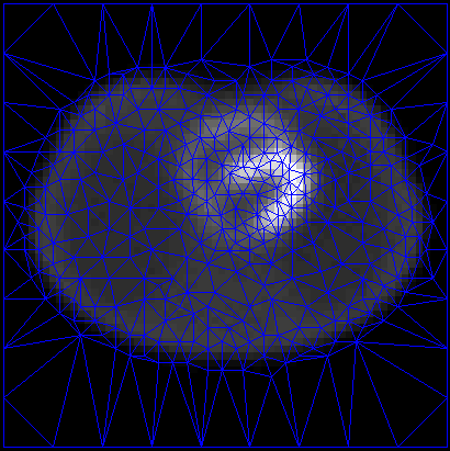
Motion-Compensated 4D Processing of Gated SPECT Perfusion Studies In this paper we present two 4D motion-compensated reconstruction algorithms for gated cardiac SPECT. The algorithms are based on a deformable content adaptive mesh model (CAMM), a compact image representation that is well suited for motion tracking of non-rigid objects like the heart. In this paper, we modify our previous approaches to preserve the brightening of the myocardium with wall thickening. Although this brightening is actually an artifact of the partial volume effect (PVE), it is an important diagnostic feature in clinical practice, and can be diminished by 4D reconstruction. Our experiments show that the new algorithms produce image sequences that closely match the expected intensity variations at the spatial resolutions of interest. In addition it is shown that the proposed method outperforms methods that do not take into account PVE. |
|
|
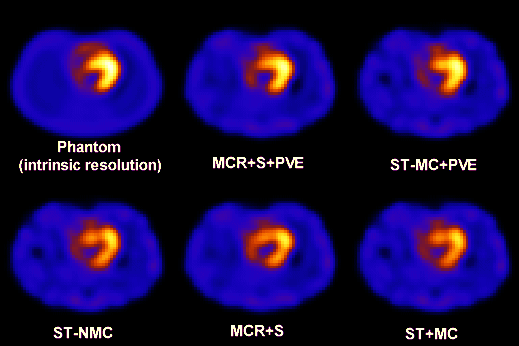 We compared the proposed reconstruction method (MCR+S+PVE) with inter-iteration smoothing (MCR+S+PVE) with a deformable mesh method that does not work to preserve myocardial brightening (MCR+S). We also compared the proposed pixel-based motion-compensated filter (ST-MC+PVE) with a version that does not preserve myocardial brightening (ST-MC), and with a simple temporal smoothing that is not motion-compensated (ST-NMC). The same spatial filter (order 5, cutoff=0.5) and temporal filter (g=1) were applied in each method. These images are used clinically for the express purpose of wall motion and thickening assessments, which can only be seen in a cine display. For more details send a request to: jovan [at] brankov [dot] com |