|
4D
smoothing of gated SPECT images We present a new 4D smoothing method for cardiacgated SPECT. The method uses motion-compensated spatiotemporal filtering, based on motion estimated using a global leftventricular shape model, and a deformable mesh, which accounts for myocardial brightening. The high noise levels in gated SPECT can cause difficulties for motion estimation methods that are based on local information, such as optical-flow techniques. In the proposed approach, we mitigate these problems by using a global shape model of the left ventricle (LV). This model is used to generate a volumetric mesh which is deformed from frame to frame to track myocardium motion through time. As opposed to surface based motion estimation, which follow the motion trajectories of points on the LV surface, our approach is a volumetric method, which aims to follow points within the myocardium. Therefore the method is explicitly designed to track and maintain the relative position of myocardial tissue elements. In addition, the proposed motion estimation allows incorporation of myocardial brightening, which is an artifact of the partial volume artifact, but plays a useful role in clinical assessment of wall thickening. Clinical SPECT data are used in this preliminary study to demonstrate the proposed method.
4D Smoothing of gated SPECT images using a left-ventricle shape model and a
deformable mesh For more details send a request to: jovan [at] brankov [dot] com
Deformable
mesh model
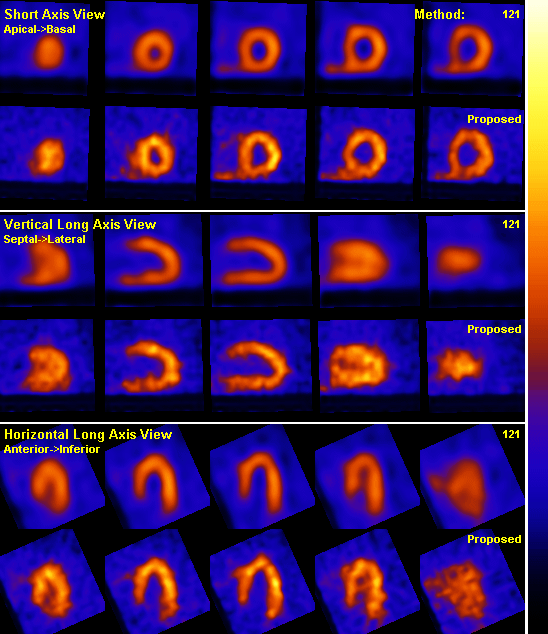
Patient
: male |
||||||||||||||||||||||||||||||||||||||||||||||||||||||
|
|
||||||||||||||||||||||||||||||||||||||||||||||||||||||
|
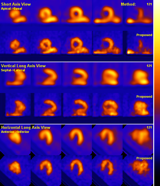
Patient
: female Note: Scroll down for second patient.
| ||||||||||||||||||||||||||||||||||||||||||||||||||||||
|
| ||||||||||||||||||||||||||||||||||||||||||||||||||||||
|
© 2004 brankov.com |
||||||||||||||||||||||||||||||||||||||||||||||||||||||